

GENERAL HOSPITAL
HANOI - VIENTIANE

 Tiếng Việt
 ຈັນລາວ
 English
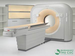
CT scanner CT-64
CATEGORY

CT scanner CT-64
I. SUMMARY of CT (Computed Tomography)
1. History of generations of computer tomography machines in the world
- In 1972, G.N Hounsfield introduced the first computerized tomography (CLVT) machine in the UK. It takes 72 hours for one photogram.
- In 1987, the first generation of helical CT scanners (spiral CT scanners) was born to help reproduce images on vertical and horizontal layers, instead of only in cross-sectional layers. Take about 1 second / 1 cut layer.
- In 2003, the first 64-row CT scanner was introduced, to help reproducing images below 1mm.
- In 2005, the first 2-source CT scanner was produced.
- In 2008, the second generation of 2-source CT scanner (defenition flash) was introduced.
- Currently, the 128, 256 and 320 series machines have been introduced (launched in July 2011, 108 hospital installed in February 2012).
2. Principle of CT machine: The main component of a CT machine is an X-ray tube and detectors, which are placed in a circular machine cavity (gantry) in opposite positions and can rotates around the patient's body. The X-ray tube is considered the "heart" of the machine. A very narrow beam of X-rays comes from the ball, passes through part of the body and is picked up by the receiver. This receiver consists of one or more arrays of receivers, made up of scintillation crystals or ionization chambers, that allow quantification of the attenuation of X-rays after passing through the body. The sensitivity of the receivers is much higher than that of X-ray film. The computer system converts this quantified information into an image. Any organ or organization of the body that has a high level of radiation resistance (bones, teeth, stones, calcifications, bleeding ...) will be white and vice versa if it is low radiation, it will be dark (fat, fluid, lungs, gas...).
 Figure 1: The principle of a CT machine with 64 receiver arrays, the width of a receiver array is 0.5 mm, the length of the whole detectors is 32 mm.

The Brilliance 2 and 64-slice CT Scanner by Philips at diagnostic imaging.
3. Spiral CT
The term “helical CT” (helical or spiral CT) is used to refer to CT machines that can scan in a spiral mode. Up to now, all CT machines can simultaneously scan in two modes: axial and spiral cutting. Shaft cutting is when the ball rotates, the table moves step by step and the ball will beam when the table stops moving. Axis cutting mode is often used for radiotherapy, GammaKnife and CyberKnife techniques for the purpose of high-precision post-capture images, unaffected by patient movement. The disadvantage of this mode is that shooting is slow, following each step of the table movement, starting from the top to the bottom of the examination organ, the outer edge of the 2D or 3D rendering has a ladder shape (Figure 3). Spiral cutting is when the ball rotates and emits rays, the table moves continuously, the trajectory of the ball relative to the patient's body is a spiral, similar to the peeling of a grapefruit (Figure 2). The advantage of spiral cutting is that the shooting speed is faster, the noise caused by movement (respiration, peristalsis, etc.) can be overcome, and the image boundaries are built continuously, without distortion.

Spiral CT scanning.
|
|
B |


These are the CT images of the face of two different subjects. A: CT scan in axial mode, the bone boundary on the 3D image has a ladder shape (white arrow). B: CT scan in spiral cutting mode, high quality 3D rendering, bone border is quite continuous, just like real image.
4. CT multi-array probe
Currently in the world, CT machine manufacturers have stopped producing single-slice CT or receiver array. The machine with the lowest configuration currently is the dual-slice CT and in the near future will only produce CT with four or more receivers. CT manufacturing technology develops in two directions: (1) Increasing the number of receiver arrays to speed up capture, for example, Philips, Siemens, GE have launched CT 128 receiver arrays, Toshiba has CT 320 rows. The fastest CT scanner available today allows imaging of the heart and coronary vessels in one beat, imaging of the entire brain and cerebral perfusion in one revolution of the balloon (0.35 seconds), (2) Increasing the number of beam sources ( from one source to two sources), this type of camera allows shooting with two different energy levels, producing two types of images and can then be superimposed.
If based on the number of receiver arrays that classify CT machines, there will be many types. However, according to Fergus V.Coakly and Bonnie N. Joe, Department of Diagnostic Imaging, University of San Francisco, California, USA, CT machines are divided into the following 3 main groups, based on clinical application:
CT with 4 rows of receivers (including 6, 8 rows): Used to take pictures of all common diseases in the head, face, neck, chest, abdomen and extremities. 4-sequence CT is still used for angiography, but the image quality is not as high as 16 or 64 row CT.
16-row CT receiver: Used to scan for all common pathologies, in addition to predominately for non-cardiac blood vessels.
CT ≥64 receiver array: Used for all applications of CT thanks to its high speed, especially advantageous in diagnosing heart and coronary diseases, imaging moving organs such as lungs, gastrointestinal tract, imaging perfusion (brain, liver, kidney)... or patients with multiple injuries (need to take snapshot, many organs at once and the patient struggles). To capture the heart and coronary arteries, the machine needs to choose two overlapping times to take pictures: (1) The time when the heart stops beating (diastolic phase), (2) The time when the contrast material is maximally absorbed into the heart and pulse. rim.
|
|
|
|
|
A: 3D image of the abdominal aorta, taken with a 16-row CT machine |
B: 64 probe CT images - tetralogy of Fallot |


The comparison of images taken on a 16-row CT and 64 receiver array.
II. INDICATIONS CT 64 LINES
1. Indications for CT thoracic CT
- Aortic aneurysm, coarctation of the aorta
- Pulmonary infarction
- Aortic dissection
- Triple rule out protocol: only one imaging program helps to exclude pulmonary infarction, coronary artery occlusion and thoracic aortic dissection
- Other diseases of the lung, mediastinum, chest wall, ... (such as conventional CT)
2. Indicated in brain damage
- Early cerebral ischemia
- Perfusion of brain tumors
- Vascular malformations...
- Other craniocerebral diseases such as conventional CT scan: hemorrhage, inflammation, tumor,... especially the volume of hematoma in the brain parenchyma can be calculated.
3. In the evaluation of vascular diseases: detecting abnormalities and vascular lesions of the whole body from the skull to the blood vessels of the upper and lower extremities on both sides.
· Vascular malformations
· Aneurysm
· Narrow or blocked arteries → Especially for stenotic lesions, the degree of stenosis can be calculated by automatic calculation program “circulation” and specialized processing software syngovia.
4. Indications for CT scan of 64 rows of coronary arteries:
* Adults: coronary angiography can be performed in all cases of different heart rhythms except complete arrhythmias, atrial fibrillation, ventricular fibrillation, etc.
- Suspected coronary syndrome (moderate and low risk)
- Assess coronary status in patients who are afraid of angiography by traditional methods
- After a stress test, the clinical results are disproportionate
- After coronary artery bypass surgery, evaluate the bridge (bypass graft)
- After angioplasty or coronary stenting
- Follow up the lesions on patients who have had coronary angiography before
- Annual follow-up of patients after heart transplant
- Evaluation of congenital coronary abnormalities
* Children: Suspected congenital heart disease, coronary artery abnormalities such as coronary fistula, origin abnormalities, Kawasaki disease, etc.
5. Indications for 64-sequence CT in congenital heart disease: The complex congenital heart such as tetralogy of Fallot, right ventricular double outlet, complex aortopulmonary collateral circulation, pulmonary artery aplasia, toxic left ventricle most, coarctation of the aorta, aortic rupture, coronary aneurysm, and ductus arteriosus.
6. Other indications like conventional CVT
General advantages: very fast survey time
· Cranial: injuries, inflammation, tumor, ...
Chest: masses in the lungs, mediastinum, pulmonary fibrosis, dilated PQ...
· Abdomen: hepatobiliary disease (tumor, trauma, obstructive jaundice,...) pancreas, spleen, two kidneys, bladder...; diseases of the gastrointestinal tract (inflammation, tumors, etc.), intra-abdominal tumors
Pelvic organs: tumors of the ovaries, uterus, ...
· CT scan in ORL: sinusitis, maxillofacial tumor, arch..., CT scan of stony bones: trauma, inflammation, spongy fibrosis, etc.
· CT eye: orbital tumors - posterior eyeball, FCC, protrusion CRNN...
III. CONTRAINDICATIONS
- General contraindications related to contrast agents.
+ Patients with chronic renal failure (cannot conduct dialysis after the scan), severe liver function impairment.
+ Severe allergic reaction to contrast
High fever and dehydration.
- Pregnant patients, especially in the first 3 months (during the embryonic stage, young, dividing cells are very sensitive to X-rays, if X-ray or CT scan during this period can cause abnormalities to appear). fetal defects.)
- Patients at high risk must undergo cardiac catheterization immediately after angiography
- Very large coronary calcification (total calcification score >1000 points) due to image noise of calcium
- Complete arrhythmia.
IV. THE LIMITATION OF CT
Because of the strong penetrating power of X-rays, CT is more difficult to detect soft lesions than MRI.
CT is difficult to detect damage to articular cartilage (hip, knee, ankle, shoulder, elbow, wrist...) and ligaments. CT is of low value in diagnosing spinal cord injury.
Due to operating on the principle of densitometry, organs with the same density and located next to each other will be difficult to distinguish boundaries on CT images, for example, metastatic cancer nodes are mixed in the soft tissue or other lesions. Injury to the neck is located in the muscle.
The image resolution of CT is not as high as that of MRI, especially for software organization, so it is difficult for CT to diagnose small lesions, such as scattered fibrous lesions in the brain, brainstem infarction. , brain damage in diabetes... Brain damage in the base of the skull is also difficult to diagnose due to artifact.
The patient is subjected to radiation exposure to X-rays. The use of these techniques for MSCT is much higher than for conventional X-rays. For example, radiation exposure when taking a routine chest X-ray film, the patient receives a radiation dose of 0.1 mSv, while a single CT scan ≥ 64 sequences, the radiation dose can be up to 5 mSv, ie. 50 times more than conventional X-ray. There are now reports that women who have previously had a 64-slice CT angiogram have a higher risk of breast cancer than the general population.
According to estimates by the Royal College of Radiologists in Australia and New Zealand, the cancer rate increases by 0.04% for patients receiving a 64-slice CT scan once a year.
V. SOME LIMITATIONS OF 64 LINES CT IN CLASSIFICATION OF HEART AND CIRCULATION
Due to the long shadow rotation time (330 milliseconds), the temporal resolution is long (165-200 milliseconds) so the survey time is long: 8-10 cardiac leads (RR interval) with a fasting time. Breathe for 8-12 seconds.
In order to obtain a good coronary picture, the average heart rate is <65 beats/min, so anti-arrhythmic drugs must be used → cannot be captured with contraindications to beta-blocker anti-arrhythmic drugs, cases of high heart rate after taking anti-arrhythmic drugs.
Image noise due to movement of the heart, of respiration.
High irradiation dose: from 8-25 mSv.
Use multiple contrast agents: to increase drug concentration, limit evaluation of small vessels (especially distal coronary artery branches <1.5 mm).
SOME IMAGES OF MSCT


Image of Sigma colon tumor metastasis in the Axial, Cronal, and sagital planes (T3N1Mx)


The MSCT Image of lower extremity blood vessels. The image of the urinary tract reconstructed on MSCT